
Cornea & Dry Eye Care
The clear front window of your eye, looked after by people who do only this
The cornea is the transparent dome at the very front of your eye. It carries about two-thirds of your focusing power, and it has no blood supply to help it heal quickly. When it thins, scars, dries or gets infected, vision changes fast. Most of what we treat here is very fixable, provided it is caught before the damage sets.
What the cornea does, and why it matters so much
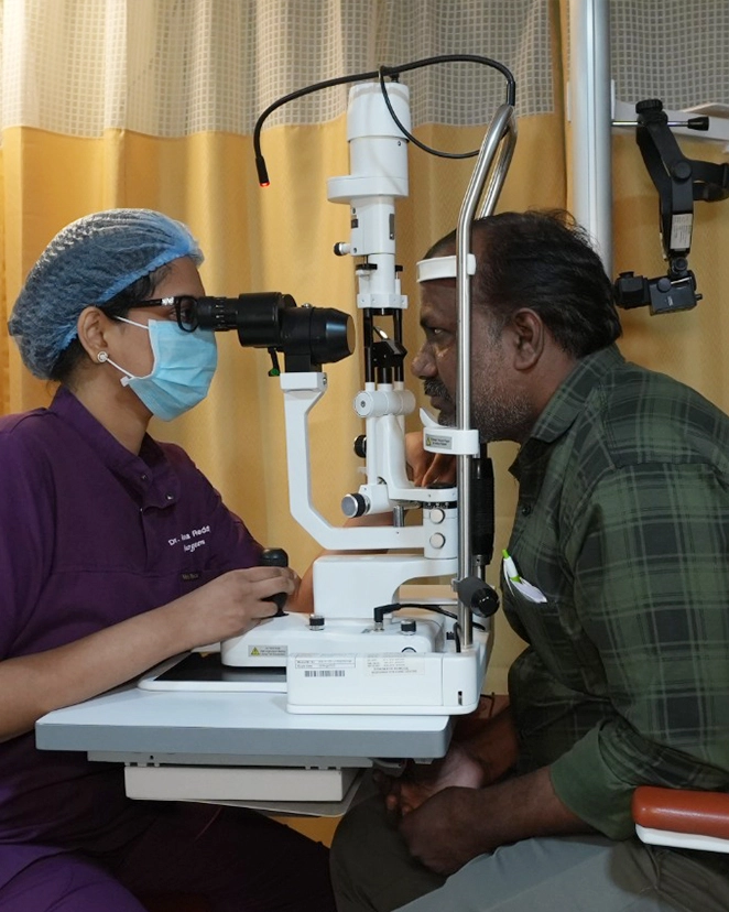
Hold a finger just in front of your eye and the clear curved surface it almost touches is the cornea: half a millimetre of tissue, five distinct layers, and no blood vessels at all. It stays clear precisely because it has no blood supply, drawing what it needs from tears on the outside and fluid on the inside. That is also why it heals slowly and why a small infection can turn into a permanent scar within days.
Because the cornea bends light before the lens ever gets a chance to, its shape decides how sharp your vision is. A cornea that is thinning and bulging, scarred, swollen or simply too dry on the surface will blur vision no matter how good your retina is. Corneal problems are also the ones people most often mistake for "just tiredness" or "power change" and live with for years.
Not sure if this applies to you?
A single examination answers it definitively. If you do not need treatment, we will tell you that too — and when to come back.
Why your eyes burn, sting and feel gritty
Dry eye is rarely a lack of water alone. In most people the oil layer of the tear film has failed, so tears evaporate before they can do their job. That distinction changes the entire treatment.
Meibomian gland dysfunction
Around 30 small oil glands in each lid seal the tear film so it does not evaporate. When they block or thicken, tears dry off in seconds. This causes the majority of dry eye we see, and plain lubricating drops do very little for it.
Blepharitis
Chronic inflammation and debris along the lash line, sometimes with mites or bacterial overgrowth. Lids look red and crusted in the morning, eyes feel worse on waking, and the irritation keeps returning until the lid margin itself is treated.
Screens and reduced blinking
Your blink rate falls by more than half while reading a screen, and incomplete blinks leave the lower cornea exposed. This is why symptoms peak on working days and settle over a holiday, and why it now affects people in their twenties.
Aqueous deficiency
The tear glands genuinely produce too little fluid. Often linked to Sjogren syndrome, rheumatoid arthritis, thyroid disease or long-term diabetes, so we look for the systemic cause rather than only treating the surface.
Hormones and medication
Dryness rises sharply around and after menopause. Antihistamines, antidepressants, beta blockers, isotretinoin and some blood pressure tablets also reduce tear production. We check your medicine list before blaming the eye.
Environment and lens wear
Air conditioning, ceiling fans overhead at night, dust, two-wheeler commuting without protection and long contact lens hours all speed up evaporation. Hyderabad summers make every one of these worse.
An honest word: in most people dry eye is controlled rather than cured. What good treatment achieves is comfortable eyes, stable vision and a healthy surface with a routine you can actually keep up. Anyone promising a permanent one-visit cure is selling something.
Treatment matched to the cause, not to the symptom
We measure tear break-up time, tear volume and gland structure first, then treat what the tests show. Two people with identical symptoms often need completely different treatment.
- Preservative-free lubricantsThe right viscosity for your tear film, in preservative-free form. Preservatives in cheap bottles used more than four times a day become part of the problem, which is why some people feel worse the longer they use drops.
- Warm compresses and lid hygieneSustained warmth at around 40 degrees for ten minutes melts thickened oil, followed by lid margin cleaning. Unglamorous, done nightly, and the single most effective thing most patients can do for themselves.
- Meibomian gland expressionIn clinic, the blocked glands are warmed and expressed directly. It relieves the load that home compresses cannot shift, and is usually repeated over a few sittings rather than once.
- Punctal plugsTiny silicone or collagen plugs close the tear drainage openings so the tears you do make stay on the eye longer. Useful in true aqueous deficiency, reversible, and fitted in a few minutes without anaesthetic injection.
- Topical cyclosporine or lifitegrastAnti-inflammatory drops that treat the inflammation driving the dryness rather than just wetting the surface. They sting for the first two weeks and take two to three months to show benefit, so we tell you that upfront.
- Omega-3 and dietRegular omega-3 intake improves the quality of gland oil in some patients. The evidence is modest, not dramatic, so we suggest it as support alongside treatment rather than as the treatment itself.
- Intense pulsed light (IPL) therapyPulses of light applied to the skin below the lids reduce inflammation and liquefy stubborn gland secretions. Given as a course of three to four sessions, it works best for evaporative dry eye and rosacea-related lid disease.
- Scleral contact lensesFor severe dry eye, the lens vaults over the cornea and holds a reservoir of fluid against it all day. It is also our main option for restoring vision in irregular or scarred corneas without surgery.
Steroid drops settle a bad flare quickly, but used unsupervised for weeks they raise eye pressure and can bring on cataract. If a chemist has been refilling a steroid drop for you, bring the bottle and let us check your pressure.
When the cornea thins and starts to bulge
Keratoconus usually begins in the teens or early twenties, when the cornea gradually weakens and pushes forward into a cone shape. Caught early it can be stopped. Caught late it may need a transplant, and that gap is entirely a matter of timing.
How it shows up
Vision blurs and distorts in a way glasses cannot quite fix. The classic pattern is a prescription that keeps changing, rising astigmatism at every visit, ghosting or streaking around lights at night, and a young patient who is told each time that their power has simply increased again.
Eye rubbing matters more than people think
Hard, habitual rubbing is strongly linked to progression, and most young patients with keratoconus also have allergic eye disease that makes them rub. Treating the allergy and genuinely stopping the rubbing is part of the treatment, not a side note.
Cross-linking (C3R or CXL) halts progression
Riboflavin drops are absorbed into the cornea and activated with controlled ultraviolet light, creating new bonds between collagen fibres so the tissue stiffens. It takes under an hour, is done under anaesthetic drops, and stabilises the cornea in the large majority of eyes.
What cross-linking does not do
It stops the cornea getting worse. It does not undo the distortion already there, so most patients still need glasses, rigid or scleral contact lenses after it. That is the honest trade: you keep the vision you have rather than gaining new vision.
When vision needs more than lenses
If contact lenses no longer fit or the cornea has scarred, intracorneal ring segments can flatten the cone, and a phakic lens implant can correct residual power in a stable eye. Transplant is the last option, not the first, and fewer than one in ten treated eyes reach it.
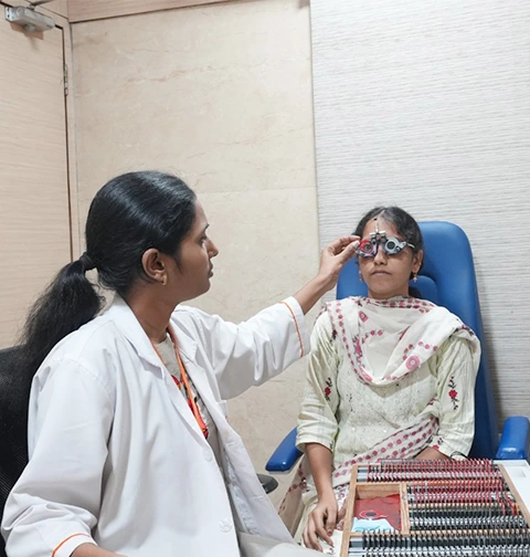
If keratoconus runs in your family, or you had it diagnosed in one eye, the other eye needs topography every six to twelve months until it is confirmed stable. Progression is silent until vision has already dropped.
Replacing only the layer that has failed
Modern corneal surgery rarely replaces the whole cornea. We replace the diseased layer and leave your healthy tissue in place, which means faster recovery, stronger eyes and far less rejection risk.
| Procedure | What is replaced | Usually done for | Visual recovery |
|---|---|---|---|
| DALK (deep anterior lamellar keratoplasty) | The front and middle layers; your own inner lining is kept | Advanced keratoconus, scars that spare the inner layer | 3 to 6 months, with sutures out over the first year |
| DSEK / DSAEK (endothelial keratoplasty) | Only the thin inner lining and its membrane | Corneal swelling, Fuchs dystrophy, damage after old cataract surgery | 4 to 12 weeks, often with a single small incision |
| PKP (penetrating keratoplasty) | The full thickness of the central cornea | Deep scarring, failed grafts, severe infection or perforation | 6 to 18 months; glasses or a rigid lens usually needed after |
| Therapeutic / patch graft | A section of cornea or sclera, to seal and save the eye | A perforated ulcer or an eye at immediate risk | Aimed at saving the eye first; optical grafting can follow later |
Donor tissue comes from a registered eye bank and is screened before use. Waiting time depends on availability, and we will tell you honestly where you stand rather than book a date we cannot keep. Rejection is possible at any point in the life of a graft, which is why lifelong follow-up and knowing the warning signs matter as much as the surgery itself.
The fleshy growth that creeps across the eye
A pterygium is a wedge of thickened conjunctiva that grows from the corner of the eye onto the cornea. It is very common in this part of India, and it is not a cancer.
Why it is so common here
Years of ultraviolet light, dust and dry wind drive it. Farmers, construction and roadside workers, drivers and daily two-wheeler commuters are the people we see most often, usually from their thirties onward.
What it feels like
Redness that never fully settles, a gritty foreign body sensation, and cosmetic concern. As it advances onto the cornea it pulls the surface out of shape, causing astigmatism and blurring long before it reaches the pupil.
When it should be removed
When it is growing towards the visual axis, when it induces significant astigmatism, when it stays inflamed despite treatment, or when it restricts eye movement. A small quiet pterygium can simply be watched with sun protection and lubricants.
How we remove it
The growth is excised and the bare area covered with a graft of your own conjunctiva, secured with fibrin glue or fine sutures. It takes around 30 minutes under local anaesthetic, and the eye is uncomfortable and red for about a week.
Recurrence, plainly stated
Simple excision without a graft recurs in a large share of cases, sometimes more aggressively than the original. With a conjunctival autograft recurrence falls to a small percentage, but it is never zero, and sunglasses afterwards genuinely reduce the risk.
The rest of what a cornea service handles
Some of these are emergencies. If you are reading this with a painful red eye and blurred vision, call us now on +91 40-4245 6666 rather than finishing the page.
Corneal ulcer and infection
An infected ulcer can scar the visual axis within days. We scrape and culture to identify whether it is bacterial, fungal or amoebic, then treat with intensive targeted drops. Fungal ulcers after injury with crop matter or a tree branch are common in our region and need weeks of treatment.
Allergic eye disease
Vernal keratoconjunctivitis makes children itch, rub and become intensely light sensitive, mainly in summer. Left untreated the rubbing damages the cornea and is a well-recognised route into keratoconus, so we treat it properly rather than handing over an antihistamine.
Corneal and scleral tear repair
Penetrating injuries from grinding, glass, firecrackers or road accidents are repaired in theatre, often the same night. Getting the eye closed early and correctly decides how much vision can be salvaged later, sometimes with a second procedure.
Chemical injury
Lime, acid, detergent or industrial chemical splashes need water irrigation immediately, at the site, for at least fifteen minutes before travelling. Do not stop to find an eye wash. Alkali burns are the more dangerous and the more deceptive of the two.
Recurrent corneal erosion
A sharp pain on opening the eyes in the morning, months or years after a scratch from a fingernail or paper edge. The healed surface has not bonded properly and lifts again. Treated with lubricating ointment, a bandage lens or a small surface procedure.
Corneal swelling and dystrophies
Fuchs dystrophy and swelling after older cataract surgery make vision worst in the morning and clearer as the day goes on. Early cases are managed medically; when it progresses, endothelial keratoplasty replaces just the failed inner layer.
Where waiting genuinely costs you vision
Some eye conditions can safely be watched. Several of these cannot, because the damage they cause does not reverse once it has happened.
Keratoconus only moves one way
Cross-linking stops progression but cannot restore a shape already lost. Every month of untreated progression in a teenager is thickness and clarity that will not come back.
Lost oil glands do not regrow
Meibography shows glands that have dropped out permanently. Treating dysfunction early preserves the glands you still have, which is the whole point of coming in before symptoms are severe.
An ulcer scars in days, not months
A central scar from a treated-too-late infection is a lifelong drop in vision that may then need a transplant. Contact lens wearers with a painful red eye should be seen the same day.
Pterygium is easier to remove early
Once it crosses the pupil it has already distorted the cornea, and even after successful removal some scarring and astigmatism can remain.
Chronic dryness damages the surface
Years of untreated dryness cause surface breakdown, fluctuating vision and unreliable measurements, which then complicates cataract and refractive surgery when you need it.
How often your cornea should be checked
| Your situation | Recommended check-up |
|---|---|
| Regular contact lens wearer | Every year, and same day if the eye turns painful or red |
| Keratoconus, still progressing | Topography every 6 months until confirmed stable |
| Keratoconus, stable after cross-linking | Once a year, lifelong |
| Teenager with allergic eye disease and heavy rubbing | Topography at diagnosis, then yearly |
| Dry eye under treatment | Every 4 to 6 months, sooner if drops stop working |
| After a corneal transplant | Week 1, then monthly for 6 months, then lifelong yearly review |
| Outdoor work with sun and dust exposure | Every 2 years, or when a growth becomes visible |
The equipment behind the diagnosis
A recommendation is only as good as the measurement it rests on. These are the machines your findings come from.
 Corneal Topography & Tomography
Maps corneal shape and thickness to detect keratoconus early
Corneal Topography & Tomography
Maps corneal shape and thickness to detect keratoconus early
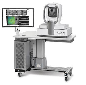 Anterior Segment OCT
Cross-sectional imaging of corneal layers, scars and grafts
Anterior Segment OCT
Cross-sectional imaging of corneal layers, scars and grafts
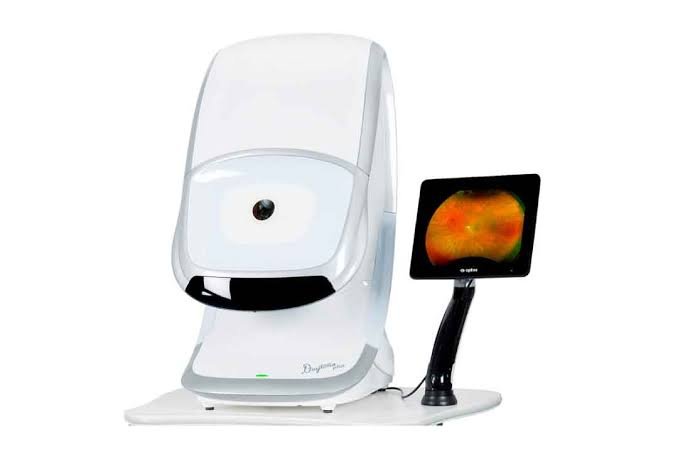 Meibography & Tear Film Analysis
Photographs the oil glands and measures tear break-up time
Meibography & Tear Film Analysis
Photographs the oil glands and measures tear break-up time
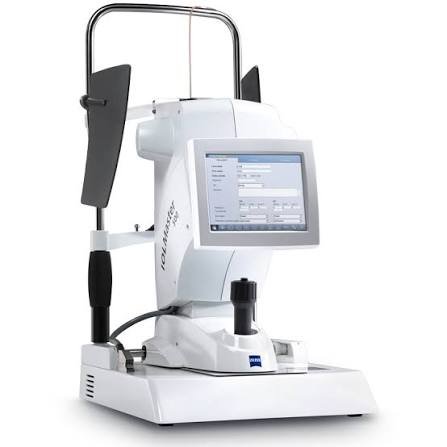 Specular Microscopy
Counts endothelial cells before surgery or transplantation
Specular Microscopy
Counts endothelial cells before surgery or transplantation
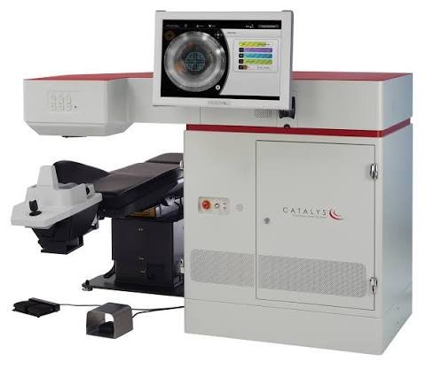 Corneal Cross-Linking System
Controlled UV-A with riboflavin to stiffen a thinning cornea
Corneal Cross-Linking System
Controlled UV-A with riboflavin to stiffen a thinning cornea
Step by step, from first visit to final review
No surprises. Here is exactly how this unfolds.
-
Detailed surface and corneal examination
Vision, refraction and a slit-lamp examination with dye staining to see exactly where the surface is breaking down. For dry eye we measure tear break-up time and tear volume rather than working from your description alone.
-
Imaging and measurement
Topography and tomography map the shape and thickness of your cornea, ASOCT looks through its layers, and meibography photographs your oil glands. These numbers are what tells us whether keratoconus is progressing or holding steady.
-
Explanation with your own scans on screen
You see your gland images and corneal maps, and we explain what they mean in plain language, including what treatment will not achieve. If the answer is lid hygiene and better habits rather than a procedure, we will say so.
-
Treatment plan and costs in writing
Drops, in-clinic procedures, cross-linking or surgery, with a written estimate and the insurance position before anything is booked. For transplants we also explain donor tissue availability and realistic timing.
-
The procedure
Cross-linking, IPL sessions, punctal plugs and pterygium surgery are day-care procedures under anaesthetic drops or local anaesthetic. Transplants are planned admissions, and we walk you through the day before you arrive.
-
Follow-up that actually continues
Dry eye is reviewed at four to six months to see whether the plan is holding. Cross-linked eyes are re-imaged to confirm stability. Grafts are followed lifelong, because early rejection treated within days usually settles and late rejection often does not.
Why patients choose us for cornea
A dedicated dry eye clinic
Not a drops prescription at the end of a general consultation. Tear film testing, meibography, gland expression and IPL under one roof, with a plan reviewed over months.
Cornea specialists, not generalists
Keratoconus, transplantation and ocular surface disease are handled by consultants who do this work regularly, which matters most in the cases that are not straightforward.
NABH-accredited theatres
Independently audited sterilisation and infection control across all centres. In corneal surgery, where infection risk decides the outcome, that is not a formality.
24x7 emergency cover
Corneal injuries, chemical burns and acute infections do not keep clinic hours. Call +91 40-4245 6666 and someone competent will see you.
Five centres, one standard
The same consultants, protocols and follow-up whether you come to our Masab Tank flagship or a district centre, with 6,25,000+ patients treated since 1993.
Where cornea is available
Listed plainly so you do not travel to the wrong centre.
| Centre | Status | What is offered |
|---|---|---|
| Masab Tank, Hyderabad | Available | C3R cross-linking, DALK, DSEK, PKP, IPL dry-eye therapy |
| KPHB, Kukatpally | Available | Dry eye, keratoconus and cross-linking |
| Warangal | Referred | Corneal transplants and cross-linking are co-managed with Masab Tank |
| Nalgonda | Referred | Corneal transplants and cross-linking are performed at Masab Tank |
| Badvel, Kadapa | Referred | Corneal transplants and cross-linking are performed at Masab Tank |
Cornea & Dry Eye — your questions answered
How is your dry eye treatment different from the drops I buy at the pharmacy?
A pharmacy drop wets the surface for twenty minutes and treats nothing. We first measure why your eyes are dry: whether the oil glands have blocked, whether tear production has genuinely dropped, or whether inflammation is driving it. Treatment then follows that finding, which might be gland expression and IPL, anti-inflammatory drops, punctal plugs, or a change to your existing medication. Many people who have been buying drops for years find their actual problem was never a lack of lubricant.
Can keratoconus be treated without a transplant?
In the large majority of cases, yes. Cross-linking stops the cornea from getting worse, and vision is then managed with glasses, rigid gas-permeable or scleral contact lenses. Ring segments or a phakic lens implant help selected eyes. Transplantation is reserved for corneas that have scarred or become too irregular for any lens to fit, and the earlier you are diagnosed, the less likely that is to be you.
How long does recovery take after a corneal transplant?
It depends on which layer was replaced. After endothelial keratoplasty such as DSEK, most people see a clear improvement within four to twelve weeks. After a full-thickness PKP, vision improves gradually over six to eighteen months as the sutures are removed, and glasses or a rigid contact lens are usually needed at the end. Drops continue for a long period in every case, and follow-up does not stop once the eye feels normal.
Can I have LASIK if I have dry eye?
Sometimes, but not before the dryness is treated. LASIK temporarily reduces corneal nerve sensation and makes dry eye worse for several months, so operating on an untreated dry eye reliably produces an unhappy patient. We treat the surface first, re-measure, and then decide. If dryness is severe, or if topography shows any suspicion of keratoconus, we will advise against laser altogether and discuss an implantable lens instead.
What causes a pterygium, and will it come back after surgery?
Long-term exposure to ultraviolet light, dust and dry wind is the main cause, which is why it is so common among people who work or commute outdoors here. Removal with a conjunctival autograft brings the recurrence rate down to a small percentage, compared with a high rate after simple excision alone. It is not zero, and recurrence is more likely in younger patients and those who go straight back to full sun exposure without protective glasses.
Can children get keratoconus or dry eye?
Yes, and keratoconus in children tends to progress faster than in adults, so it is treated more urgently once confirmed. The pattern to watch for is a child with allergic eye disease who rubs their eyes constantly and needs a new spectacle power every few months. Dry eye in children is usually screen-related or allergy-related rather than glandular, and it responds well to blinking habits, screen breaks and treating the allergy properly.
How do I know whether it is dry eye or something else?
You often cannot, because dry eye, allergy, blepharitis, an early corneal infection and a raised eye pressure can all begin as redness and discomfort. The signs that it is not simple dryness are pain rather than irritation, vision that drops, light sensitivity that stops you opening the eye, or symptoms in one eye only. Any of those need examination the same day, not another bottle of drops.
My eyes water all the time. How can that be dry eye?
It is one of the most common patterns we see. When the surface dries out it becomes irritated, and irritation triggers a flood of reflex tears that are watery, poor quality and drain away immediately. So the eye waters and stays dry at the same time. The other possibility is a blocked tear duct, which we check for, because the treatment for that is completely different.
Where does donor corneal tissue come from, and can it be rejected?
From registered eye banks, donated after death by consent, and screened for infection before use. Rejection is possible at any point in the life of a graft, though it is much less likely with partial-thickness procedures such as DSEK or DALK than with a full-thickness transplant. The warning signs are redness, pain, light sensitivity and a drop in vision. Reported within a day or two, most rejection episodes are reversed with intensive drops.
Is any of this covered by insurance?
Corneal transplantation, pterygium surgery and repair of corneal or scleral injuries are covered by most health policies and by the government schemes we are empanelled for. Cross-linking is covered by many insurers but not all, and dry eye treatment, IPL sessions and specialty contact lenses are generally out-of-pocket outpatient costs. Our insurance desk checks your specific policy and tells you the exact figure before you commit to anything.
Often looked at alongside this
LASIK & Refractive Surgery
Freedom from glasses through LASIK, SILK, PRK, ICL or refractive lens exchange — matched to your cornea, not to a trend.
Learn moreSILK Laser Vision Correction
Flapless laser vision correction on the ELITA platform — no blade, no flap, minimal dry eye.
Learn moreComprehensive Ophthalmology
A full diagnostic examination that checks the whole eye — not just what prescription you need.
Learn moreCataract Surgery
Micro-incision and femtosecond laser-assisted cataract surgery, with the full range of monofocal, toric and multifocal lenses.
Learn moreGet your cornea and tear film measured properly
One examination separates simple dryness from something that needs treating now. If your eyes are only tired, we will tell you that and send you home.