
Retina Care
The part of the eye that fails quietly, and the checks that catch it in time
The retina rarely warns you. Diabetic damage, a leaking macula, even a tear at the edge can sit there for months while your vision reads 6/6 on the chart. Which is why retina care is built around scanning, not around symptoms.
What the retina does, and why damage stays silent
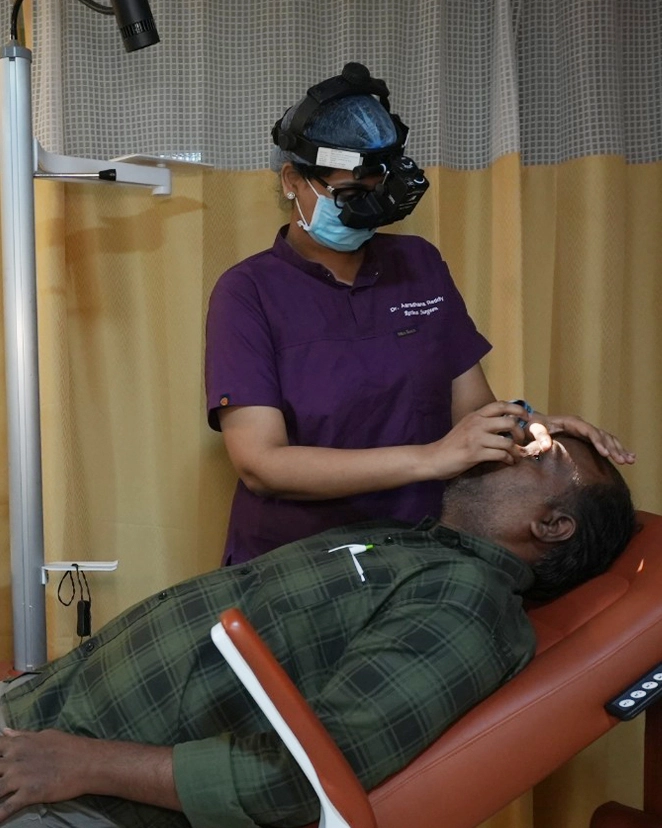
The retina is a layer of light-sensitive tissue about as thick as a sheet of paper, lining the back of the eye. It converts light into signals the brain reads as sight. At its centre sits the macula, a patch a few millimetres across that handles everything needing detail: faces, reading, the number on an autorickshaw.
The retina has no pain fibres. A tear, a leak or a slow build-up of fluid produces no ache, no redness, nothing you can feel. Worse, if one eye is affected the other quietly compensates, so the loss is only noticed when the good eye is covered by accident. By the time a person says "my vision dropped last week", the scan often shows changes that have been present for a year or more.
Most of what we treat here cannot be reversed once the macula is scarred. It can, however, be caught early and held stable for decades. That is the whole argument for annual screening, and it is not a marketing line.
Not sure if this applies to you?
A single examination answers it definitively. If you do not need treatment, we will tell you that too — and when to come back.
Diabetic retinopathy
Diabetes damages the smallest blood vessels in the body first, and the retina is where the smallest ones are. Duration of diabetes matters more than how high your sugar went last week.
Why diabetes reaches the retina
Years of raised blood sugar weaken the walls of the retinal capillaries. Some balloon and leak fluid or blood; others close off entirely, leaving patches of retina starved of oxygen. Both processes run silently, which is why the length of time you have had diabetes predicts retinopathy better than any single sugar reading.
Non-proliferative retinopathy (NPDR)
The early stage. The scan shows microaneurysms, small dot haemorrhages and yellow exudates, but vision is usually still normal. Nothing needs to be injected or lasered at this point. What it needs is documented monitoring, and serious work on sugar, blood pressure and cholesterol, which genuinely slows the progression.
Proliferative retinopathy (PDR)
The oxygen-starved retina starts growing fragile new vessels. They bleed into the vitreous, and the scar tissue that follows can pull the retina off the eye wall. This stage is sight-threatening and treatable, usually with pan-retinal laser, sometimes with injections or vitrectomy surgery. It should not be watched and waited on.
Diabetic macular oedema
Fluid collecting at the macula is the commonest reason a person with diabetes loses reading vision, and it can occur at any stage, including mild NPDR. It shows up on an OCT scan as measurable thickening long before you notice blur. Anti-VEGF injections are the first-line treatment, with focal laser or a steroid implant in selected eyes.
What annual screening actually involves
Dilating drops, a dilated examination, an OCT scan of the macula and a wide-field retinal photograph. Fluorescein angiography only if the other tests raise a question. Budget forty minutes, and do not plan to drive yourself home, as the dilation blurs near vision for three to four hours.
What treatment can and cannot do
Laser and injections are far better at preserving vision than at restoring it. Vision already lost to a scarred macula usually does not come back. This is an uncomfortable thing to hear and the honest reason we push screening so hard: the treatment works best in eyes that still see well.
If you have diabetes, you need a dilated retina check every year even when your vision is perfect and your sugars are controlled. A normal reading on the eye chart does not rule out retinopathy.
Age-related macular degeneration
AMD affects the macula alone. Side vision stays, so people rarely go completely blind, but the central detail needed for reading and recognising faces is exactly what it takes.
Dry AMD
The slow form, and about nine in ten cases. Yellow deposits called drusen build up under the macula and the tissue thins over years. There is no treatment that reverses it. AREDS2-formula supplements slow progression in intermediate cases, and stopping smoking helps more than any tablet.
Wet AMD
Abnormal vessels grow under the retina and leak fluid or blood. Vision can distort or drop over days, not years. Straight lines bend, and a grey or blank patch appears in the centre. This is the form where weeks of delay change the final outcome.
The Amsler grid
A printed square grid you look at from reading distance, one eye at a time, every week. If lines wave, blur or go missing where they were straight last week, that is your cue to call us rather than wait for the next appointment. We give you the chart and show you how to use it.
Anti-VEGF for wet AMD
An injection into the eye blocks the protein driving the leak. Typically three monthly doses to start, then a treat-and-extend schedule guided by OCT. Most eyes hold their vision and many improve, but this is control rather than cure, and injections often continue for years.
What raises your risk
Age above sixty, smoking, a parent or sibling with AMD, high blood pressure, and a diet short on leafy greens and fish. Smoking is the one modifiable factor with the largest effect, and stopping reduces risk even after decades.
Why timing decides the result
Once leaked fluid gives way to scar tissue at the macula, no injection recovers that vision. An eye treated in the first weeks of wet AMD usually keeps useful reading vision. The same eye treated six months on usually does not.
Retinal detachment and tears
This is one of the few genuine eye emergencies. The retina peels away from its blood supply, and every day it stays detached costs recoverable vision.
- A sudden shower of floatersNot the one or two specks you have had for years. A fresh cloud of dots, a cobweb, or what people describe as soot or pepper appearing over a few hours often means blood or pigment released by a tear.
- Flashes of lightBrief arcs or streaks in the side vision, most obvious in a dark room or when you move your eyes. They come from the vitreous gel tugging on the retina, which is the moment a tear can form.
- A curtain or shadowA dark area coming in from one side, top or bottom, that does not clear when you blink. This means the retina has already lifted. Come in the same day, not the next.
- A sudden drop in vision in one eyePainless, sudden and one-sided is the pattern that should worry you. Pain is not part of it, so the absence of pain is no reassurance at all.
- Who is at higher riskHigh myopia above roughly -6 dioptres, a previous detachment in either eye, blunt injury to the eye, lattice degeneration seen on a past examination, and eyes that have had complicated cataract surgery. If this is you, ask for a peripheral retina check every year.
- What happens when you reach usDilation and an indirect examination of the full peripheral retina, with scleral indentation to see the very edge, plus an ultrasound scan if blood blocks the view. A tear alone can be sealed with laser the same visit. A detachment is scheduled for surgery, usually within a day or two.
Emergency line: +91 40-4245 6666. If the macula is still attached when you reach us, the odds of a good visual result are far better. Once it detaches, surgery can reattach the retina but the central vision that returns is often incomplete.
Retinal treatments, and what each one is for
Retina treatment is rarely a single event. Most plans combine one of these with regular scanning, and we will tell you at the start roughly how many visits to expect.
| Treatment | What it does | Mainly used for | What to expect |
|---|---|---|---|
| Anti-VEGF injection | Blocks the protein driving leakage and new vessel growth | Diabetic macular oedema, wet AMD, vein occlusion | A few minutes in a sterile room; three loading doses, then reviewed on OCT |
| Barrage laser | Seals healthy retina around a tear so fluid cannot get under it | Retinal tears, holes, lattice degeneration | Outpatient, about ten minutes, no incision, home immediately |
| Pan-retinal photocoagulation | Treats oxygen-starved peripheral retina to stop new vessels forming | Proliferative diabetic retinopathy | One to three sittings; some loss of night and side vision is the accepted trade |
| MIVS vitrectomy (23G / 25G) | Removes vitreous gel, blood and scar tissue through sub-millimetre ports | Vitreous haemorrhage, tractional detachment, macular hole, membranes | Day care or one night; face-down posturing for some days if gas is used |
| Scleral buckling | A silicone band indents the eye wall so the retina can settle back | Selected rhegmatogenous detachments, often in younger eyes | Slightly longer recovery; the spectacle number usually changes |
| Intravitreal steroid implant | Releases anti-inflammatory medicine inside the eye over months | Oedema that responds poorly to anti-VEGF | Fewer visits, but eye pressure and cataract must be watched |
Two honest caveats. Vitrectomy will speed up cataract formation in most eyes over fifty that still have their natural lens, so a cataract operation often follows within a year or two. And an eye that has lost central vision to long-standing scarring is unlikely to gain it back from any of the above.
Other retinal conditions
Not everything at the back of the eye is diabetes or age. These come through our clinics regularly, and several of them affect people well under forty.
Retinal vein occlusion
A retinal vein blocks, usually where an artery crosses it, and vision in one eye blurs painlessly overnight. Blood pressure, diabetes and cholesterol are the usual background, so a physician review is part of the treatment. Anti-VEGF handles the macular oedema; laser is added if new vessels appear.
Central serous chorioretinopathy
A blister of fluid under the macula, most often in men between thirty and fifty, and linked to stress and to steroid use in any form, including inhalers and skin creams. Roughly three in four settle on their own within three months. Stopping the steroid matters most; laser or photodynamic therapy is reserved for fluid that persists.
Retinopathy of prematurity
Babies born before 34 weeks or under 2,000 g can grow abnormal retinal vessels in the first weeks of life. The first screening is due between day 20 and day 30, and the treatable window is narrow, sometimes days. Treated in time with laser or anti-VEGF, most babies keep good vision; missed, ROP is a leading cause of childhood blindness in India.
Macular hole
A small full-thickness break at the exact centre of vision, causing a dark or distorted patch when you read. Vitrectomy with membrane peeling and a gas bubble closes over nine in ten holes, though it asks for face-down positioning afterwards. Holes operated within six months do considerably better than older ones.
Epiretinal membrane
A fine sheet of cells grows over the macula and wrinkles it, so straight lines look bent and print looks slightly doubled. Mild ones need nothing but an annual OCT. Surgery to peel the membrane is worth it when distortion interferes with daily tasks, and it improves vision in most but not all eyes.
The case for screening an eye that sees perfectly well
Almost everyone we treat for advanced retinal disease had a window, several years wide, when a fifteen-minute check would have changed the outcome.
The retina cannot hurt
There are no pain fibres in the retina. Tears, leaks and slow oedema are entirely painless, so waiting for a symptom means waiting past the easy stage.
A scan sees what you cannot
OCT measures macular thickness in microns. It picks up swelling months before you notice any blur, which is when treatment works best and costs least.
Scarring is permanent
Fluid can be dried up. Bleeding can be cleared. Scar tissue at the macula cannot be undone by any drug or surgery available anywhere.
Your physician is half the treatment
Sugar, blood pressure and cholesterol control measurably slows retinopathy. No injection replaces that, and we will say so plainly rather than pretend the eye is a separate organ.
Early is simpler and cheaper
A tear sealed with ten minutes of laser costs a fraction of detachment surgery, needs no theatre, and carries far less risk to your vision.
When to have your retina checked
| Your situation | Recommended check-up |
|---|---|
| Type 2 diabetes | At diagnosis, then every year |
| Type 1 diabetes | Within 5 years of diagnosis, then every year |
| Diabetes, with any retinopathy already found | Every 3 to 6 months, as advised |
| Pregnancy with diabetes | Before conception if planned, then each trimester |
| Age 60 and above | Every year, including a dilated macular check |
| High myopia above -6 dioptres | Every year, with a peripheral retina examination |
| Premature baby under 34 weeks or 2,000 g | First screening between day 20 and day 30 |
| New floaters, flashes or a shadow | Same day. Call the emergency line, do not wait for a slot |
The equipment behind the diagnosis
A recommendation is only as good as the measurement it rests on. These are the machines your findings come from.
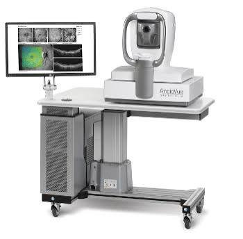 OCT & OCT Angiography
Cross-sections of the macula in microns, and vessel maps without dye
OCT & OCT Angiography
Cross-sections of the macula in microns, and vessel maps without dye
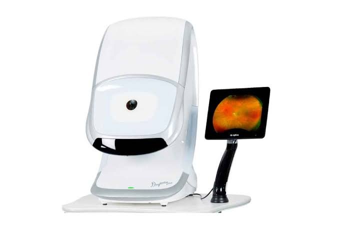 Optos Ultra-Wide-Field Imaging
Captures roughly 200 degrees of retina in a single photograph
Fundus Fluorescein Angiography
Maps leakage and closed capillaries before laser or injection
Optos Ultra-Wide-Field Imaging
Captures roughly 200 degrees of retina in a single photograph
Fundus Fluorescein Angiography
Maps leakage and closed capillaries before laser or injection
 CONSTELLATION Vision System
Micro-incision vitrectomy surgery for the back of the eye
CONSTELLATION Vision System
Micro-incision vitrectomy surgery for the back of the eye
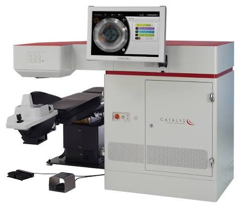 Retinal Laser Delivery Systems
Barrage laser for tears and pan-retinal treatment for diabetes
Retinal Laser Delivery Systems
Barrage laser for tears and pan-retinal treatment for diabetes
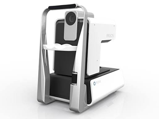 ARGOS Optical Biometer
Lens power planning when retina surgery is combined with cataract
ARGOS Optical Biometer
Lens power planning when retina surgery is combined with cataract
Step by step, from first visit to final review
No surprises. Here is exactly how this unfolds.
-
History and vision check
How long you have had diabetes, your last HbA1c, blood pressure, and what exactly you have noticed. Vision is recorded for each eye separately, because a good eye hides a bad one.
-
Dilation and retinal examination
Drops widen the pupil over about twenty minutes. The consultant then examines the full retina to its edge, with scleral indentation where a tear is suspected. Near vision stays blurred for three to four hours afterwards, so bring someone to drive.
-
OCT and wide-field imaging
Both are painless and take a few minutes. The OCT measures macular thickness against your previous scans, and the wide-field photograph gives a permanent record of the periphery to compare next year.
-
Angiography, only if it changes the plan
A dye is injected into an arm vein and photographs are taken as it fills the retinal vessels. It tints the skin and urine yellow for a day. Allergic reactions are rare but real, so we do it only when the result will alter treatment.
-
The plan, with numbers and costs
You see your own scans on the screen, with an estimate of how many injections or sittings are likely in the first year and what each costs. Insurance and Aarogyasri coverage is worked out at this point, not after.
-
Treatment and follow-up
Injections are given in a sterile theatre-grade room, laser in the clinic, surgery as day care or one night. Review is usually at four to six weeks with a repeat OCT, and the interval is then stretched as long as the scans stay stable.
Why patients choose us for retina
Vitreoretinal surgeons, not generalists
Retina work here is done by consultants who trained specifically in vitreoretinal surgery and operate on it every week, not occasionally between cataract lists.
Imaging before opinion
Nobody is told they need an injection before their OCT and fundus images are on the screen in front of them, alongside the previous scans where we have them.
Injections given under theatre discipline
Intravitreal injections carry a small but serious infection risk. We give them in a sterile setting with full aseptic protocol, which is what keeps that number small.
Costs stated before the first injection
Retina treatment is a course, not a single bill. You get the likely first-year figure in writing, including drug, procedure and follow-up scans, before you commit to anything.
Five centres, one retina protocol
Screening and follow-up happen close to home; complex vitreoretinal surgery is done at the 50-bed flagship. Same consultants, same records, no repeated tests.
Consultants who handle retina
The same consultants practise across our centres — you get the specialist, not the postcode.




Where retina is available
Listed plainly so you do not travel to the wrong centre.
| Centre | Status | What is offered |
|---|---|---|
| Masab Tank, Hyderabad | Available | Anti-VEGF injections, laser, MIVS vitrectomy, scleral buckling |
| KPHB, Kukatpally | Available | Diabetic screening, anti-VEGF injections and laser |
| Warangal | Available | Diabetic retinopathy screening, laser and intravitreal injections |
| Nalgonda | Available | Diabetic retinopathy screening, laser and intravitreal injections |
| Badvel, Kadapa | Available | Diabetic retinopathy screening and referral for treatment |
Retina Care — your questions answered
How often should someone with diabetes have a retina check?
Once a year at minimum, from the day type 2 diabetes is diagnosed, and within five years of diagnosis for type 1. If any retinopathy is already present, the interval shortens to every three to six months. In pregnancy the retina can change quickly, so screening is done before conception where possible and again each trimester.
Are intravitreal injections painful?
Almost never. The eye is numbed with anaesthetic drops or gel and cleaned with antiseptic; most people report pressure and a brief stinging from the antiseptic rather than from the needle itself. The injection takes a few seconds. Mild grittiness and a red patch on the white of the eye for a few days are common and harmless.
Does retinal detachment always cause blindness?
No, but time decides the outcome. If surgery is done while the macula is still attached, most people recover good vision. Once the macula has detached, the retina can usually still be put back but central vision often returns incomplete. Around nine in ten detachments are reattached successfully, though some need a second procedure.
What is the difference between dry and wet AMD?
Dry AMD is the slow form, where deposits build up and the macular tissue thins over years; there is no treatment that reverses it, and the mainstays are AREDS2 supplements in intermediate cases and stopping smoking. Wet AMD is when abnormal vessels grow and leak, causing distortion or loss over days to weeks. Wet AMD is treatable with anti-VEGF injections and is the reason sudden distortion should be seen quickly.
Is vitreoretinal surgery safe, and how long is the recovery?
Modern micro-incision vitrectomy uses ports under a millimetre wide, is usually done under local anaesthetic, and is day care or a single night in most cases. Vision recovers over four to eight weeks, longer if a gas bubble is used, since the bubble blurs vision until it absorbs. The real trade-offs to know about are accelerated cataract in eyes that still have their natural lens, and a small risk of raised pressure or infection.
When should a premature baby be screened for ROP?
Any baby born before 34 weeks or weighing under 2,000 g at birth needs a first retinal screening between day 20 and day 30 of life, and earlier if the baby needed prolonged oxygen. The window in which ROP can be treated is narrow, sometimes a matter of days, and there are no outward signs a parent can notice. This one appointment is not worth postponing for any reason.
Can retina treatment be done along with cataract surgery?
Yes, and often the sequence matters more than the combination. Where the retina can be seen well enough, we usually stabilise it first, because clearer media makes laser and injections more effective. Where a dense cataract blocks the view of the retina, the cataract is removed first or both are combined in one sitting. If retinopathy or AMD is present we generally advise a monofocal lens rather than a multifocal one, since multifocals reduce contrast in an eye that has little to spare.
Will one injection fix the problem?
Rarely. Anti-VEGF drugs control leakage rather than cure the underlying disease, so the standard approach is three monthly loading doses followed by review, then a treat-and-extend schedule guided by your OCT. Some eyes stretch to four-monthly and stop; others need injections for years. We will tell you honestly which pattern your scans suggest rather than promise a fixed number.
Are floaters always dangerous?
No. Most floaters come from the vitreous gel shrinking with age, are harmless, and become less noticeable over months. What matters is the change: a sudden shower of new floaters, flashes of light, or a shadow in the side vision needs a dilated examination the same day, because those three together are how a retinal tear announces itself.
Is retina treatment covered by insurance?
Vitreoretinal surgery is covered by most health insurance policies and by CGHS, ECHS, Aarogyasri and Ayushman Bharat. Intravitreal injections are covered by many policies but not all, and some cover the drug only for specific conditions, so the answer depends on your exact policy. Our insurance desk checks the wording and gives you the out-of-pocket figure before treatment begins.
Often looked at alongside this
Comprehensive Ophthalmology
A full diagnostic examination that checks the whole eye — not just what prescription you need.
Learn moreGlaucoma Treatment
The vision glaucoma takes does not come back — which is why detection, not treatment, is the real work.
Learn moreCataract Surgery
Micro-incision and femtosecond laser-assisted cataract surgery, with the full range of monofocal, toric and multifocal lenses.
Learn morePaediatric Ophthalmology
Children rarely say their vision is poor — they adapt. We find what they will not tell you.
Learn moreGet your retina looked at before it asks for attention
A dilated check with an OCT scan takes about thirty minutes and tells you exactly where you stand. If everything is normal, we will say so and tell you when to come back.