
Oculoplasty
Surgery around the eye, judged first on how it works
Oculoplasty covers the eyelids, the tear drainage system and the bony socket. Most of this work is functional: a lid that will not lift, lashes scraping the cornea, an eye that waters all day, a socket under pressure from thyroid disease. Appearance usually improves as well, but that is the by-product, not the reason to operate.
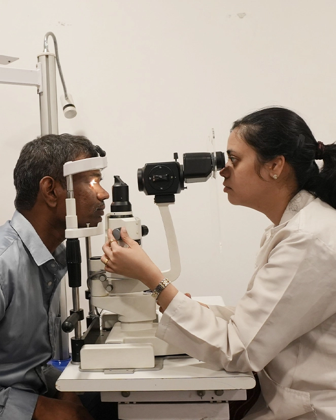
What oculoplasty actually covers
The eye sits in a bony socket, is protected by two lids that blink roughly fifteen thousand times a day, and drains its tears through a narrow channel into the nose. When any of that machinery fails, the eye itself can be perfectly healthy and still not work properly. Oculoplasty is the sub-speciality that repairs those surrounding structures.
It is easy to confuse with cosmetic surgery, because the operations happen on the face. The distinction is straightforward: we operate when a problem blocks vision, endangers the cornea, causes constant watering or infection, or follows an injury or a tumour. Where a request is genuinely cosmetic, we will say so plainly, and so will your insurer.
Not sure if this applies to you?
A single examination answers it definitively. If you do not need treatment, we will tell you that too — and when to come back.
Ptosis, and why the cause decides the operation
Ptosis is an upper lid sitting lower than it should. The right operation depends on how much of your levator muscle still works, so the cause has to be settled before anything is booked. In children there is a clock running too: a lid covering the pupil stops that eye learning to see, and that is amblyopia.
| Type | What is behind it | How it usually presents | Usual approach |
|---|---|---|---|
| Congenital | The levator muscle did not develop normally before birth | Present from infancy, often one eye, with a poorly formed lid crease | Levator resection, or a frontalis sling when muscle function is very poor |
| Aponeurotic (age-related) | The tendon of the levator stretches or slips off the lid plate | Gradual droop after middle age, a high lid crease, worse by evening | Levator advancement, usually under local anaesthetic |
| Neurogenic | A nerve problem such as third nerve palsy or Horner syndrome | Often sudden, may come with double vision or an unequal pupil | Find the cause first; surgery only once the picture is stable |
| Myogenic | Muscle disease such as myasthenia gravis or a mitochondrial myopathy | A droop that varies through the day or worsens with fatigue | Medical treatment first; surgery is cautious and sometimes avoided |
| Mechanical | Weight on the lid from a lump, scarring or chronic swelling | One heavy lid, often with a visible mass or thickened skin | Treat the cause, then reassess the lid height |
A droop that appears suddenly, comes with double vision, sits alongside an unequal pupil, or fluctuates through the day is not routine ptosis. Those need a neurological work-up before anyone reaches for a surgical date.
Entropion and ectropion
Both are mechanical problems of lid position, both are usually correctable in a single sitting, and both do quiet damage if they are tolerated for years.
Entropion: the lid rolls inward
The lid margin turns in and the lashes sweep across the cornea with every blink. It feels like grit that never washes out. Left long enough it scars the cornea, and a scar over the pupil does not come back with surgery.
Ectropion: the lid falls outward
The lower lid sags away from the eye, so tears no longer reach the drainage opening. The result is constant watering, a red raw inner lid, and a lower cornea left exposed and drying.
Why they happen
Most cases are age-related laxity of the lid tissues. Others follow scarring from burns, chronic infection or earlier surgery, or a facial nerve palsy that leaves the lid unable to close.
What the surgery does
A short day-care procedure under local anaesthetic tightens the lid horizontally and repositions the retractors so the margin sits flat against the eye again. Stitches come out at about a week.
Drops and taping are a holding measure
Lubricants and lid taping relieve symptoms and protect the cornea while you wait for a slot. They do not change lid position. If the cornea is already staining on examination, waiting is not a neutral choice.
Eyelid tumours and the signs that matter
Most lid lumps are harmless: a stye, a chalazion, a cyst, a skin tag. A small number are skin cancers, and on the eyelid these are usually curable when caught early. These are the features that move a lump from watching to biopsy.
- It bleeds or breaks downRepeated bleeding, crusting, or a centre that ulcerates and will not heal is the single most useful warning sign on a lid.
- Lashes are missing over itEyelashes do not grow through a tumour. A bald patch on the lid margin matters even when the lump itself looks small.
- It keeps growingA chalazion settles or stays put. Anything enlarging steadily over months needs a tissue diagnosis, not another course of ointment.
- The lid margin is distortedAn edge that has lost its clean line, or lashes pointing in odd directions, suggests something growing beneath the surface.
- A chalazion that returns to the same spotSebaceous gland carcinoma imitates a chalazion almost exactly. A lump recurring in the same place after drainage should go for histopathology.
- Hard, fixed or numbA lump tethered to the deeper tissue, or numbness of the skin around it, points to more than a cyst.
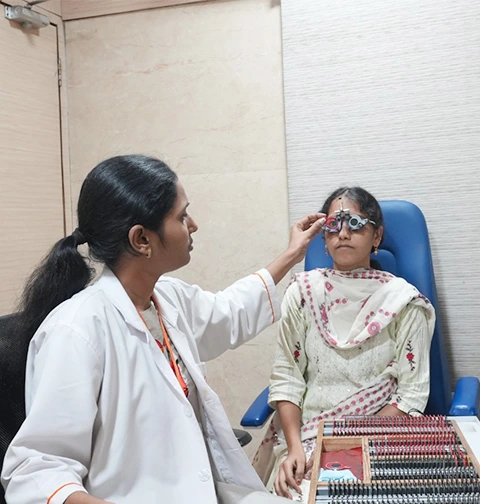
Removal is only half the operation. Taking a malignant lesion off a lid margin leaves a defect that must be rebuilt so the lid still closes, still holds tears against the eye and still protects the cornea. We plan the reconstruction before the first cut, and margins are confirmed on histopathology before we call it finished.
Eyelid, tear duct and orbital repair
Road accidents, falls, sport and workplace injuries account for most of what we repair. Timing genuinely matters here: some of this is far easier in the first few days than in the first few months.
Eyelid laceration
A cut through the lid margin has to be realigned exactly, layer by layer. A millimetre of misalignment leaves a permanent notch, a lid that never quite closes, and a patch of cornea that dries out at that spot.
Lacrimal (canalicular) laceration
Any cut on the inner third of the lid may have divided the tear drainage channel. Repaired within a few days over a fine silicone stent it usually heals open. Missed, it often means lifelong watering and a bigger operation to put right later.
Orbital fractures
A blow to the eye can break the thin floor of the socket. Not every fracture needs surgery. We operate for muscle trapped in the fracture with restricted eye movement, for double vision that is not settling, or for an eye sinking backwards. The rest we watch, with a review date.
What to do on the day
Shield the eye with a clean cup or pad taped over it, do not press on it, do not wash it out with anything, and come in. Our emergency line is +91 40-4245 6666 and runs round the clock.
When the socket is the problem, not the eye
Thyroid eye disease inflames the muscles and fat inside the orbit, pushing the eye forward. It runs in two phases, and most of the disappointments in managing it come from operating in the wrong one.
Active phase: settle it, do not cut
Redness, swelling, aching behind the eye and measurements that change from month to month mean the disease is still active. This phase is managed medically and can last a year or longer. Operating into an active orbit invites a result that shifts afterwards.
What actually helps early
Stopping smoking makes the largest single difference, larger than most drugs. Beyond that: getting the thyroid itself stable, selenium in mild disease, lubricants for exposure, and steroids or targeted therapy for moderate to severe cases.
The one true emergency
Pressure on the optic nerve at the back of the socket causes falling vision, washed-out colours and an abnormal pupil response. That needs urgent treatment, and sometimes urgent decompression, whichever phase the disease is in.
Inactive phase: surgery in strict order
Decompression first to make room in the socket, squint surgery second, eyelid surgery last. That order is not tradition. Each stage changes the measurements for the next, so doing the lids first usually means doing them twice.
Patience is part of the treatment
We generally want the disease quiet and the measurements steady for about six months before planning rehabilitative surgery. It is a long path, and we would rather tell you so at the start than halfway through.
Blocked tear ducts, from newborns to adults
A constantly watering eye is more often a plumbing problem than a tear problem. Where the blockage sits decides everything about how it is treated, so the first job is to find the level.
| Problem | Who it affects | First step | If that does not work |
|---|---|---|---|
| Congenital nasolacrimal duct obstruction | Babies, from the first weeks of life | Crigler massage: a clean fingertip over the tear sac at the inner corner, ten firm downward strokes twice a day. Roughly nine in ten clear on their own within the first year | Probing under a short general anaesthetic, usually between 9 and 12 months |
| Punctal stenosis | Adults, often alongside chronic lid inflammation | Treat the lid disease first, then a small punctoplasty in clinic | Intubation with a fine silicone stent |
| Acute dacryocystitis | Adults, occasionally infants | Same-day review, oral antibiotics and warm compresses for the infection | Drain the abscess, then DCR once it settles, otherwise it returns |
| Chronic dacryocystitis or established duct block | Adults, more often women over 40 | No drop or tablet clears an established block | DCR, either external or endoscopic endonasal |
| Watering with a clear drainage system | Any age | Look for the real cause: dry eye, lid malposition, allergy or blepharitis | Treat that cause. Operating on an open duct helps nobody |
External DCR reaches the tear sac through a small incision beside the nose. It gives the widest exposure and heals to a fine line most people struggle to find after a few months. Endoscopic endonasal DCR works entirely through the nostril, so there is no skin scar at all and the tear pump muscle is left undisturbed, but it needs good nasal access and is harder work in an unusual nose or after a failed previous DCR. Success rates are broadly comparable in experienced hands, so the choice rests on your anatomy rather than on fashion.
What waiting actually costs
Very little in oculoplasty is an emergency on the day. But several of these problems do quiet damage while they are being put up with, and a few change the outcome entirely if they are missed.
A child with ptosis has a deadline
A lid covering the pupil blocks the developing visual pathway. Amblyopia sets in early and becomes much harder to reverse after seven or eight years of age, which is why we assess a droopy lid in infancy rather than waiting for school.
Lashes rub, then the cornea scars
Entropion and a lid that will not close are uncomfortable long before they are dangerous, but the endpoint is corneal scarring or an ulcer. Once the scar sits over the pupil, fixing the lid no longer restores the vision.
Lid cancers are curable when small
A basal cell carcinoma removed early leaves a lid that still works normally. The same tumour after two years of ointment may need a far larger reconstruction, and a few types spread beyond the lid.
Thyroid eye disease can threaten the nerve
Falling vision or faded colours in an inflamed orbit means the optic nerve is under pressure. Treated promptly it usually recovers. Left for weeks, some of that loss becomes permanent.
A cut tear duct has a short window
A canalicular laceration repaired within the first few days usually heals with drainage intact. Attempted months later, the results are considerably less reliable.
When to come in, and how soon
| What you have noticed | When to be seen |
|---|---|
| Droopy lid in a baby or young child | Within weeks, before it affects visual development |
| Gradual droop in an adult, nothing else changed | At your convenience, but get it measured |
| Sudden droop, double vision or an unequal pupil | Same day. This is a neurological presentation |
| Watering eye, no discharge, no pain | Routine appointment |
| Red, swollen, tender lump at the inner corner | Same day. That is an infected tear sac |
| Lid lump that bleeds, grows or has lost its lashes | Within a week, for examination and possible biopsy |
| Bulging or aching eyes with thyroid disease | Within weeks, sooner if vision or colours are changing |
| Any injury around the eye | Immediately. Emergency line +91 40-4245 6666 |
The equipment behind the diagnosis
A recommendation is only as good as the measurement it rests on. These are the machines your findings come from.
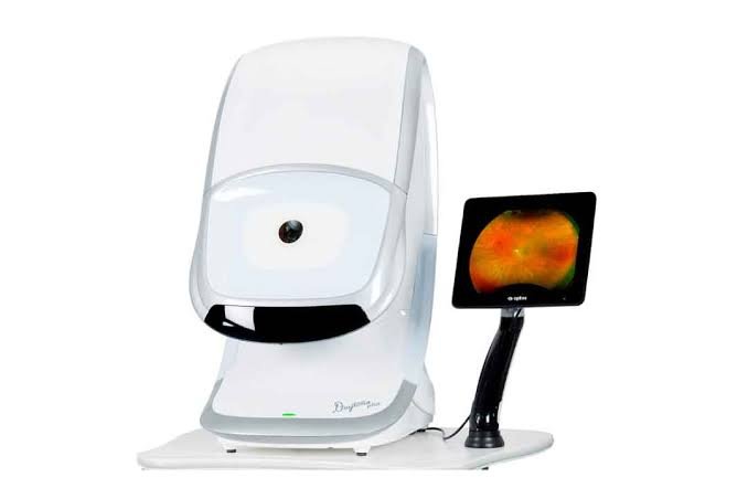 Orbital CT and MRI imaging
Maps fractures, tumours and the crowded thyroid orbit
Orbital CT and MRI imaging
Maps fractures, tumours and the crowded thyroid orbit
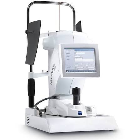 Slit-lamp with photodocumentation
Records lid measurements and lesions to compare over time
Slit-lamp with photodocumentation
Records lid measurements and lesions to compare over time
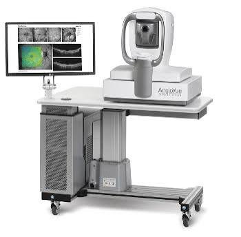 Visual field and optic nerve assessment
Documents functional field loss and detects nerve compression
Visual field and optic nerve assessment
Documents functional field loss and detects nerve compression
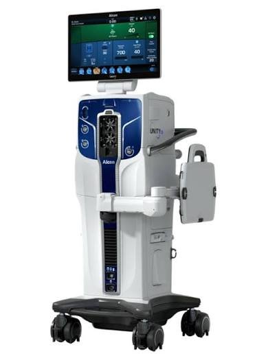 Endoscopic DCR system
Creates a new tear drainage path through the nose, with no skin incision
Endoscopic DCR system
Creates a new tear drainage path through the nose, with no skin incision
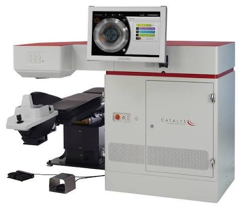 Surgical microscope
Magnified repair of lid margins and tear canaliculi
Surgical microscope
Magnified repair of lid margins and tear canaliculi
Step by step, from first visit to final review
No surprises. Here is exactly how this unfolds.
-
Consultation and measurement
Vision, proper lid measurements including margin-reflex distance and levator function, tear film and drainage assessment, and a careful look at the cornea. Photographs go on file, because lid surgery is judged against where you started.
-
Tests, only the ones that change the plan
A CT or MRI for orbital disease, trauma or a suspicious mass. Thyroid function and antibody tests where thyroid eye disease is suspected. Visual fields and optic nerve testing when the socket is crowded or an insurer needs documented field loss. Not everybody needs all of this.
-
A plan you can see, with costs in writing
What the operation will and will not achieve, which parts are functional and which are cosmetic, what your policy is likely to cover, and a full rupee estimate before anything is booked. Cashless pre-authorisation is handled by our insurance desk.
-
Surgery day
Most adult lid and lacrimal surgery is day care under local anaesthetic with light sedation. Children, orbital work, endoscopic DCR and larger reconstructions are done under general anaesthesia at our 50-bed flagship. You are usually home the same day.
-
The first two weeks
Swelling and bruising peak around day two or three and then improve steadily. Cold compresses for the first 48 hours, antibiotic ointment on the wound, skin sutures out at about a week. Most people feel presentable by the end of the second week.
-
Review, and occasionally revision
We check lid height, closure and tear drainage at one week, one month and three months, once the swelling has genuinely gone. Ptosis surgery is measured in millimetres, and a small proportion of cases need a second adjustment. We tell you that beforehand, not afterwards.
Why patients choose us for oculoplasty
Oculoplasty done by eye surgeons
These lids belong to an eye. Our surgeons are ophthalmologists first, so the cornea, tear film and vision are part of every plan rather than an afterthought.
Reconstruction planned with the excision
For lid tumours the removal and the rebuild are worked out together, and margins are confirmed on histopathology before the case is closed.
A 50-bed NABH-accredited flagship
Orbital surgery, general anaesthesia and inpatient care under one roof, with independently audited theatre and sterilisation protocols.
Joint care with squint and cornea
Thyroid eye disease and facial palsy need more than one pair of hands. Decompression, squint and lid stages are planned by the same team, in the right sequence.
Straight about functional versus cosmetic
You are told which part of your surgery an insurer should cover and which part you pay for yourself, with the estimate in writing before you decide anything.
Where oculoplasty is available
Listed plainly so you do not travel to the wrong centre.
| Centre | Status | What is offered |
|---|---|---|
| Masab Tank, Hyderabad | Available | Ptosis, thyroid eye disease, external and endoscopic DCR, trauma repair |
| KPHB, Kukatpally | Available | Eyelid and tear-duct procedures |
| Warangal | Referred | Orbital and complex eyelid surgery is co-managed with Masab Tank |
| Nalgonda | Referred | Eyelid, orbital and tear-duct surgery is performed at Masab Tank |
| Badvel, Kadapa | Referred | Eyelid and orbital surgery is performed at Masab Tank |
Oculoplasty — your questions answered
Is ptosis surgery covered by insurance?
Usually yes, when the droop is functional. If the lid blocks part of your field of vision, or you find yourself lifting your chin or eyebrow to see, most policies cover the correction. Insurers generally want documented visual field testing with the lid taped and untaped, plus clinical photographs, and we prepare that as part of the work-up. Purely cosmetic lid surgery in an eye that sees perfectly well is not covered and is paid for privately.
How long does recovery from eyelid surgery take?
Swelling and bruising peak on the second or third day and settle over one to two weeks. Skin stitches usually come out at about a week, and most people are comfortable being seen in public by the end of the second week. The final lid position is only judged at two to three months, once the last of the swelling has gone.
Will my baby's blocked tear duct clear on its own?
Most of the time, yes. Around nine in ten congenital blocks open by themselves during the first year, helped by Crigler massage: with a clean fingertip over the tear sac just below the inner corner, press firmly downwards ten times, twice a day. If the eye is still watering at nine to twelve months, or there are repeated infections, a simple probing under a short general anaesthetic clears it in most children. Bring the baby in the same day if the inner corner becomes red, swollen and tender.
What is the connection between thyroid disease and my eyes?
Thyroid eye disease is an autoimmune condition that attacks the muscles and fat inside the eye socket. It is associated with thyroid disease but not caused by the hormone levels themselves, which is why eye symptoms can appear before, during or years after the thyroid is diagnosed, and why they do not always improve once the thyroid is corrected. Getting the thyroid stable still helps. Stopping smoking helps more than any single medicine we can give you.
External or endoscopic DCR, which is better?
Neither is better in the abstract. External DCR uses a small incision beside the nose and gives the widest exposure, which matters in revision cases and unusual anatomy; the scar fades to a fine line most people cannot easily find after a few months. Endoscopic endonasal DCR is done through the nostril, so there is no skin scar and the tear pump muscle is left untouched, but it needs good nasal access. Success rates are broadly comparable, so the decision comes down to your anatomy and what has already been tried.
Is this cosmetic surgery, or does it affect my vision?
The great majority of oculoplasty is functional. Ptosis blocks the upper field of vision, entropion scars the cornea, ectropion causes constant watering and exposure, thyroid eye disease can threaten the optic nerve, and an untreated lid tumour destroys the lid. Appearance usually improves too, and that is welcome, but it is not the reason we operate. Where a request genuinely is cosmetic, we will say so plainly rather than dress it up as medical.
Is eyelid surgery safe at 70 or 80?
Age on its own is rarely the obstacle. Most lid surgery is day care under local anaesthetic, which suits older patients well, and we routinely operate on people in their eighties. What matters more is blood thinners, diabetes control, dry eye and skin quality. Aspirin or clopidogrel may need adjusting in consultation with your physician, and a very dry eye limits how much lift is safe, so occasionally we advise against surgery and treat the dryness instead.
Will I be awake during the operation?
For most adult lid and tear duct surgery, yes. Local anaesthetic with light sedation is standard, and being awake is genuinely useful in ptosis surgery, because we ask you to open and close your eyes so the height can be set on the table. Children, orbital surgery, endoscopic DCR and extensive reconstruction are done under general anaesthesia.
Will there be a visible scar?
Upper lid incisions sit inside the natural crease and are effectively invisible once healed. Lower lid work is often done from inside the lid with no skin incision at all. External DCR leaves a short line beside the nose that fades over several months, and endoscopic DCR leaves none. Reconstruction after a large tumour excision is the exception, where some scarring is unavoidable, and we will show you what to expect before you consent.
Often looked at alongside this
Comprehensive Ophthalmology
A full diagnostic examination that checks the whole eye — not just what prescription you need.
Learn moreCornea & Dry Eye
Keratoconus cross-linking, corneal transplantation and a dedicated dry-eye clinic with IPL therapy.
Learn morePaediatric Ophthalmology
Children rarely say their vision is poor — they adapt. We find what they will not tell you.
Learn moreSquint Treatment
Squint correction at any age — glasses, prisms, therapy or surgery, depending on what is actually causing the turn.
Learn moreGet the lid, the tear duct or the socket looked at properly
One examination with real measurements tells you whether this needs surgery, whether it needs watching, or whether it needs a physician rather than a surgeon.