Hospital websites list LASIK, retina and cataract as three separate services. Inside your eye they are not separate at all. The same eye that needs a spectacle number also has a retina behind it and a lens inside it, and a decision made in one of those places changes what happens in the other two. This is worth understanding, because it explains a lot of things patients find confusing: why a LASIK consultation includes a dilated retina check, why a high-myopia cataract needs extra measurements, why one person is told to wait and another is told to operate.
The three sit in a line, front to back
Light entering your eye passes through the cornea, then the natural lens, then lands on the retina. Refractive surgery reshapes the cornea. Cataract surgery replaces the lens. Retina work happens at the back, where the image is actually captured. If any one of those three is compromised, the other two cannot compensate.
That gives you a simple rule that guides most decisions we make: the retina sets the ceiling. The cornea and lens control how sharply light is delivered. The retina controls how much of that light becomes vision. You can polish the front of the eye beautifully and still get a disappointing result if the back is damaged. This is why we look at the retina first, even when the patient has come in for something else entirely.
Why a LASIK screening includes the back of your eye
Someone walks into our KPHB centre asking about laser vision correction. Their power is minus six. Reasonable request. But minus six means the eyeball is longer than average, and a longer eyeball means the retina lining it is stretched thinner than average. Thin retina is more prone to weak patches, holes and tears, particularly in the far periphery where you would never notice a problem yourself.
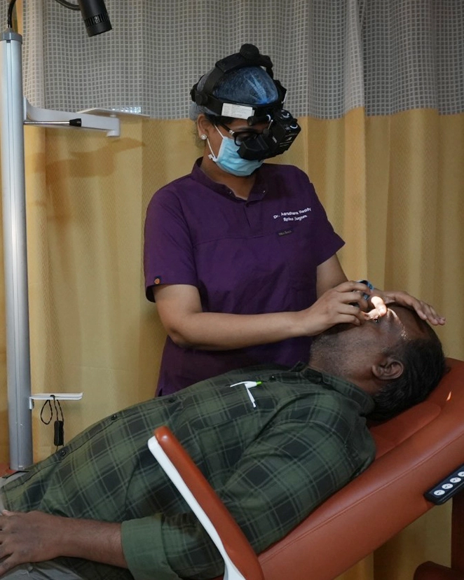
So before any laser touches the cornea, we dilate the pupil and examine the entire retina, often with ultra-widefield imaging that captures far more of the periphery than a standard fundus photo. Two outcomes are common:
- The retina is normal. We proceed with the refractive workup as planned. Most people fall here.
- There is lattice degeneration or a small hole in the periphery. We treat it with a barrier laser in the outpatient department, wait two to four weeks, then continue with the refractive plan.
Notice that in the second case the answer is almost never no. It is later. A weak spot found and sealed before surgery is a small thing. The same weak spot found after a retinal detachment is a much bigger operation with a much less certain outcome. We have written separately about what the retina examination actually involves, and it is worth reading if you are planning refractive surgery this year.
Why high myopia changes cataract surgery
Now take the same minus six eye thirty years later, with a cataract. Two things are different from a routine case.
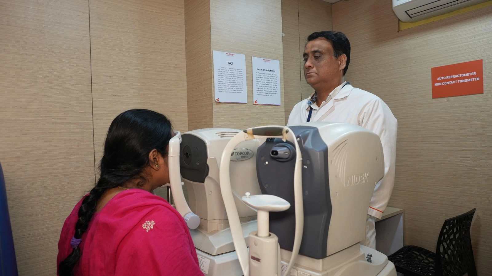
The measurements are harder. Choosing the right intraocular lens power depends on measuring the length of the eye and the curvature of the cornea very precisely. A long eye leaves less margin for error, and small measurement mistakes translate into larger refractive surprises after surgery. We use optical biometry, the IOLMaster 700 and the ARGOS biometer, precisely because they handle these eyes better than older ultrasound methods, especially when the cataract is dense.
The retina is still stretched. Cataract surgery in a highly myopic eye carries a slightly higher lifetime risk of retinal detachment than in a normal-length eye. Not high, but not zero. That is a reason to examine the retina before surgery, to counsel the patient about flashes and floaters afterwards, and to see them promptly if those symptoms appear. It is not a reason to avoid cataract surgery. A dense cataract left in place also makes it impossible to monitor the retina at all.
And if you have had LASIK before
Here is a connection that surprises many patients in their fifties. If you had laser correction in your twenties or thirties, your cornea has been permanently reshaped. When you later develop a cataract, the standard formulas used to calculate lens power assume a natural, unoperated cornea. Applied blindly, they can be off by a full dioptre or more, which means an unexpected number after an otherwise perfect surgery.
This is manageable, but only if we know. So bring your old records. The pre-LASIK spectacle power and the treatment sheet, if you still have them, are genuinely useful documents. If you do not have them, say so honestly rather than guessing, and we will use measurement methods designed for post-refractive eyes instead.
The single most useful thing a long-term spectacle wearer can do is keep one folder with every eye report they have ever received. It takes no effort and it changes surgical decisions decades later.
What this means for how you should be examined
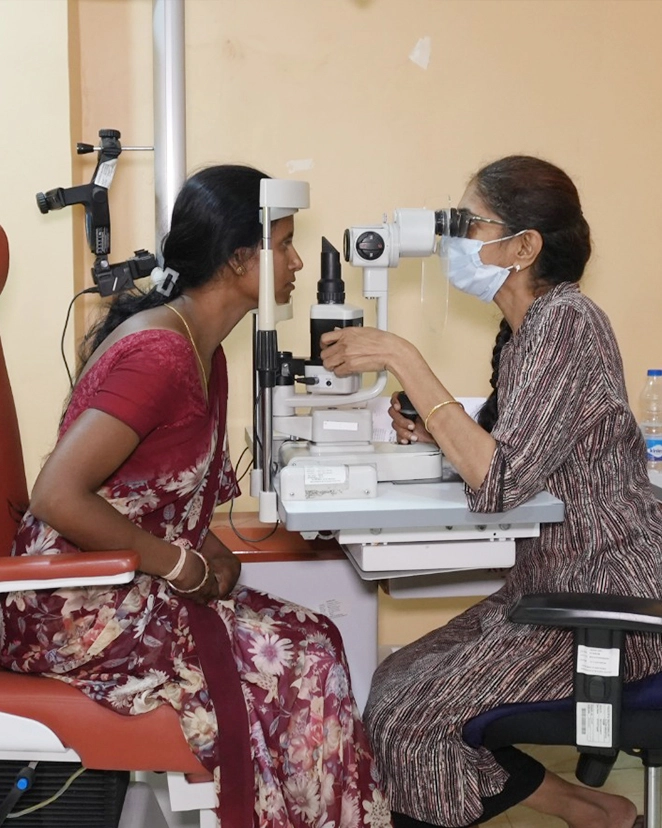
The practical implication is that a proper eye consultation is not a single-issue transaction. If you come for a LASIK opinion, you should leave knowing the condition of your retina and your intraocular pressure, not only your candidacy for laser. If you come for cataract surgery, you should leave knowing whether anything at the back of the eye will limit your result, so your expectations are set before surgery rather than adjusted after it.
This is also why we keep these specialities under one roof rather than referring patients across the city. At our Masab Tank flagship the cornea, retina, cataract and glaucoma teams see the same imaging on the same day. At KPHB, refractive and retina services run together, which matters because the majority of refractive candidates are myopic and myopic eyes need retinal attention.
None of this makes your visit longer in any meaningful way. A dilated examination adds about half an hour and leaves you light-sensitive for a few hours, which is why we ask you to bring someone who can drive you home. That is the entire cost of knowing what is actually happening in all three layers of your eye.
If you are considering any of these procedures, book a full evaluation rather than a procedure-specific consultation, and ask for the retina findings to be explained to you along with everything else. You can reach us on +91 90001 80035 or through our appointments page. Bring your old prescriptions. They matter more than you think.