Laser vision correction happens on the front surface of the eye. The retina is at the back. Patients reasonably ask why we insist on dilating the pupil and examining the retina before a procedure that never goes anywhere near it. The answer has nothing to do with the laser and everything to do with why you needed glasses in the first place.
Short-sightedness is a structural change, not just a number
Most people who come for refractive surgery are myopic. Myopia is usually caused by the eyeball growing slightly too long from front to back. The image then focuses in front of the retina instead of on it, and you see blur at distance.
Here is the part that gets left out of most explanations. When the eyeball elongates, the retina lining the inside has to stretch to cover a larger surface. It does not grow more tissue. It thins. The thinning is worst at the far periphery, which is exactly where you have almost no useful vision and would therefore never notice anything wrong.
The higher the power, the more elongation, and the more likely those thin patches become. This is a property of your eye that laser surgery does not change. Correcting minus eight to zero on the cornea makes you see beautifully. It does not shorten the eyeball and it does not thicken the retina. Your retina remains a minus eight retina for life.
What we are actually looking for
The dilated examination and widefield imaging are hunting for a short list of specific findings:
- Lattice degeneration. Patches where the retina is thinned and the underlying support is weakened. Common in myopes, usually harmless, occasionally the site where a tear begins.
- Retinal holes and tears. Small breaks in the retina. Fluid can pass through a break and lift the retina off the wall behind it, which is a retinal detachment and a surgical emergency.
- Vitreous changes. The gel filling the eye separates from the retina as we age, earlier in myopic eyes. Usually uneventful. Occasionally it pulls hard enough to create a tear.
- Anything unrelated but important. Diabetic changes, an unusual optic disc, macular thinning. A dilated exam sees all of it.
None of these produce symptoms in their early stages. You cannot feel a retinal hole. There is no pain, no blur, nothing. That is precisely why it is looked for rather than waited for.
What happens if we find something
Patients often assume a retinal finding means the refractive surgery is cancelled. It usually does not. The typical sequence is:
- A break or high-risk lattice patch is identified during the pre-operative examination.
- It is sealed with a barrier laser in the outpatient department. This takes a few minutes, is done at the slit lamp with anaesthetic drops, and feels like bright flashes rather than pain. You go home the same day.
- The laser scar takes two to four weeks to form a strong adhesion around the weak area.
- You return, the retina is re-examined, and the refractive plan proceeds.
So the practical effect is a delay of a few weeks, not a cancellation. The alternative, which is to skip the check, is that the same weak spot is discovered later as a detachment. That means an operating theatre, a longer recovery, weeks of positioning restrictions and a final visual result that depends heavily on whether the central macula was involved. The difference between those two paths is a twenty-minute examination.
The examination itself
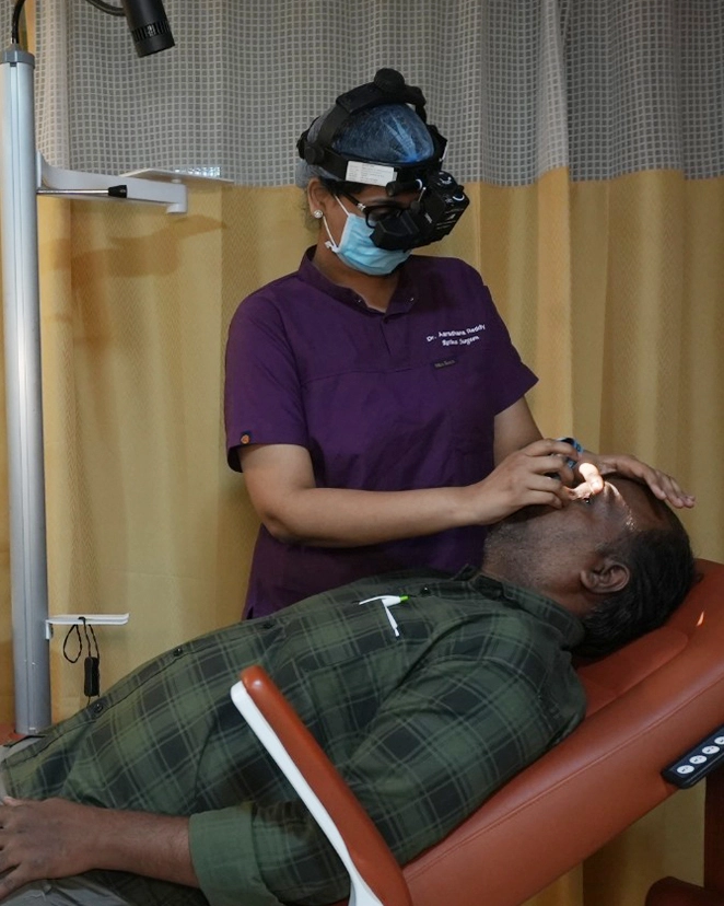
Expect drops that sting briefly and take about thirty to forty minutes to fully widen the pupil. Then an examination with an indirect ophthalmoscope, where the doctor uses a bright headlight and a hand-held lens and asks you to look in different directions to bring the far periphery into view. Some pressure may be applied on the eyelid to rotate the periphery into sight. It is not painful but it is not comfortable either.
We often add ultra-widefield imaging, which captures a very large area of the retina in a single photograph. This is useful for two reasons: it documents what your retina looked like on that date, and it makes comparison at future visits far more reliable than memory or a written description.
Afterwards your pupils stay large for four to six hours. Everything looks washed out and bright, and near vision is poor. Do not plan to drive yourself home, and bring sunglasses. If you are coming to KPHB after work, factor this into your evening.
It does not stop after surgery
This is the part most patients forget within a year of getting rid of their glasses. Once you see clearly without correction, it is easy to stop thinking of yourself as short-sighted and stop attending eye checks altogether. Your retina did not get the memo.
If you were moderately or highly myopic before laser correction, you should continue having a dilated retinal examination roughly every one to two years, and you should know the warning symptoms cold:
- A sudden shower of new floaters, particularly with a dark or cobweb appearance
- Flashes of light, most noticeable in dim surroundings or when you move your eyes
- A shadow, veil or curtain coming in from one side of your vision
Any of these is a same-day problem, not a next-week problem. Our Masab Tank centre runs a 24x7 emergency service on +91 40-4245 6666 specifically for this kind of thing. A tear treated on the day it appears is an outpatient laser. The same tear a week later may be an operation.
If you are planning laser vision correction, ask for the retinal findings to be explained to you in the consultation and note down whether lattice or any break was seen. That single line in your record is information you will want ten years from now. Our retina team works alongside the refractive clinic at both Hyderabad centres, so this is usually settled in one visit.