Most eye checks after the age of 50 take about eight minutes and end with a new spectacle prescription. That is a refraction. It is useful, and it is not an examination. The three conditions that cause the most avoidable sight loss in this age group, glaucoma, diabetic retinopathy and macular disease, all give normal-looking distance vision until they are well advanced, and none of them is detected by reading letters off a chart. Knowing what should happen in the room lets you tell one from the other.
The six parts of a real examination
- Vision tested one eye at a time, then refraction. Both eyes together hides a great deal, because the better eye covers for the worse one. The prescription that comes out of this is the by-product, not the purpose.
- Slit lamp examination of the front of the eye, including the lens. This is where an early cataract is graded, and where lid margin disease, corneal changes and a shallow anterior chamber are picked up. A shallow chamber matters, because it flags the small group at risk of angle closure glaucoma, in whom dilating drops need care and a laser can prevent an emergency.
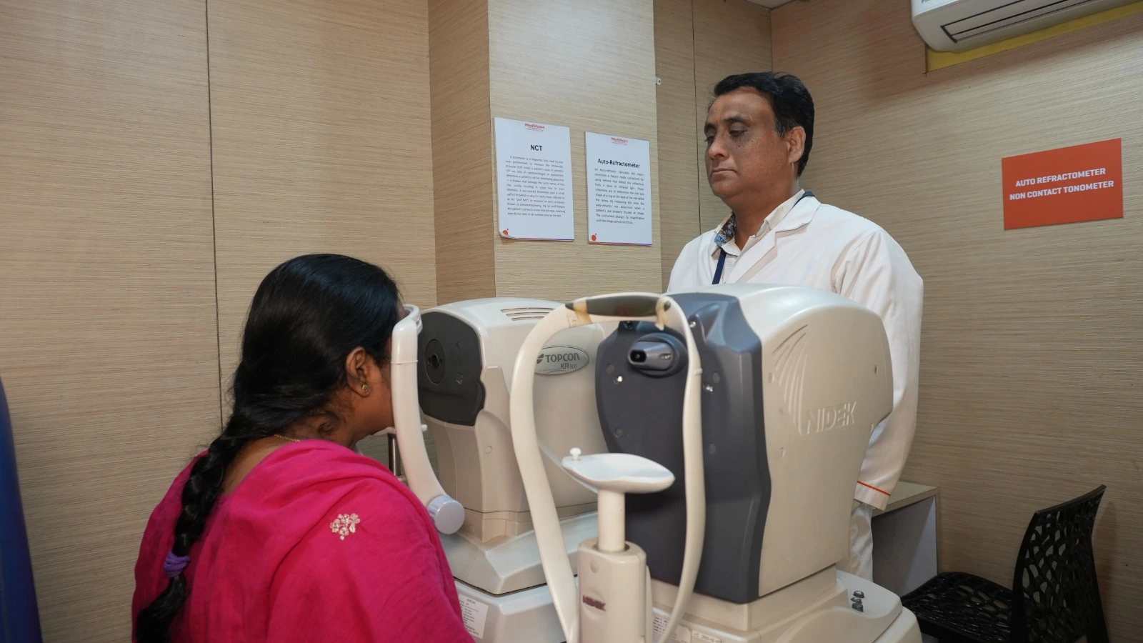
- Intraocular pressure. Quick and painless. Worth knowing that pressure alone neither confirms nor excludes glaucoma; plenty of people with glaucoma have pressures within the normal range. It is one input, not the verdict.
- A dilated examination of the retina in both eyes. The single most important part, and the part most often skipped.
- Assessment of the optic nerve head, ideally recorded, so that next year's picture can be compared with this year's. Optic nerve fibres do not grow back. Glaucoma is only caught early by noticing change over time.
- A look at the macula, with an OCT scan if anything is unclear, and an Amsler grid you can use at home for the distortion that patients notice long before a chart does.
Add one more thing that is not a test: a written record you can carry to the next visit. An eye check with nothing to compare against is worth far less than the second one in a series.
Dilation is the part people talk their way out of
Dilating drops take twenty to forty minutes to work and leave near vision blurred and light uncomfortable for three to five hours. It is genuinely inconvenient, which is why patients decline it and why some clinics do not push.
Through an undilated pupil the view of the retina is roughly like inspecting a room through a keyhole. The periphery, where retinal tears and early diabetic changes often begin, is largely invisible. Ultra-widefield imaging, which we run at several centres, captures a great deal in a single undilated photograph and is excellent for screening and for follow-up comparison. It still does not fully replace a dilated look with a lens when the history calls for one.
Practical version: come with someone who can drive, or take a cab, bring sunglasses, and do not plan detailed work for the rest of the afternoon. Once a year that is a fair trade.
What changes based on your history
The standard check above is the floor. Several things raise it.
- Diabetes. A dilated retinal examination every year from the date of diagnosis, regardless of sugar control or vision, and more often once any retinopathy appears. Diabetic macular oedema is treatable and painless, and the treatable window closes quietly. Our retina service handles this.
- A parent or sibling with glaucoma. Your risk is several times higher. Start regular checks earlier, and expect pressure, optic nerve imaging and a visual field test rather than pressure alone. See the glaucoma page for what the tests involve.
- High blood pressure, which shows in the retinal vessels and occasionally announces itself as a sudden blockage in one eye.
- High myopia, above roughly minus six. The retina is thinner and more prone to tears and detachment, so the periphery needs looking at specifically, and any new floaters or flashes need same-day attention.
- Long-term steroids, including inhalers for asthma and creams for skin conditions. They can raise eye pressure silently.
- Hydroxychloroquine for rheumatoid arthritis or lupus, which needs baseline and then periodic retinal monitoring with OCT and fields.
- Any previous eye surgery or injury, which changes what we look for and how often.
What a free screening can and cannot do
Free camps and free consultation offers are useful and we run them, but it is only fair to say where the limits are. A short screening reliably picks up a visually significant cataract, gross refractive error, an obviously raised pressure and an eye that clearly needs a closer look. It is a good filter, and it brings in people who would otherwise never walk into a hospital.
What a screening usually does not include is dilation, a visual field test, OCT imaging, or the time to build a record you can compare next year. So treat a free check as a first pass. If it says everything looks fine and you have diabetes, a family history of glaucoma or new symptoms, you still need the full examination. If it flags something, take that referral seriously rather than waiting for symptoms to arrive.
How often, and when not to wait
- 50 to 60, no risk factors, no symptoms: every one to two years.
- Over 60: annually.
- Diabetes, glaucoma in the family, high myopia, or an existing eye condition: annually at minimum, or as often as your ophthalmologist specifies, from whatever age you are now.
Ignore the schedule entirely and come the same day for any of these: sudden loss of vision, a curtain or shadow moving across the field, a sudden shower of floaters or flashes, straight lines looking bent, a painful red eye with haloes around lights, or double vision that has just started. Masab Tank runs 24-hour emergency cover for exactly these.
If it has been more than two years, book the longer appointment rather than the quick one, and say on the phone that you want a dilated check. All five of our centres do the full examination; Warangal and Nalgonda are open seven days, and our Badvel centre works with government schemes for patients for whom cost is the deciding factor.