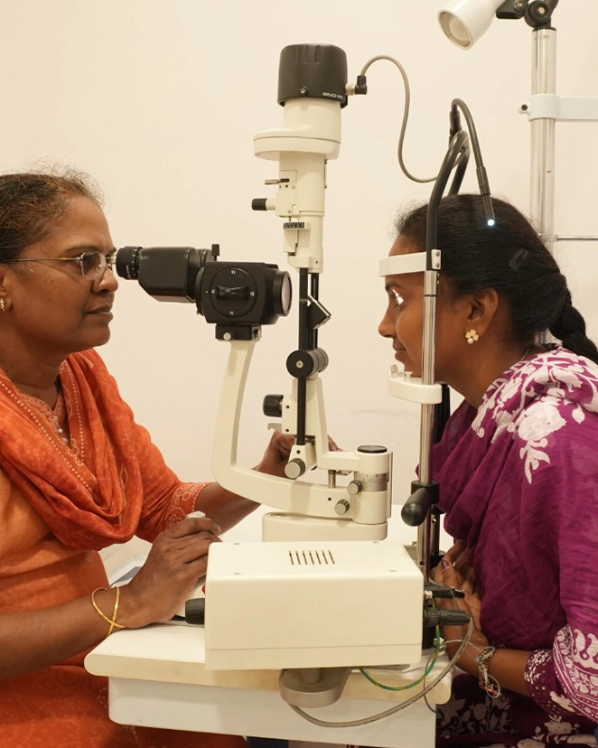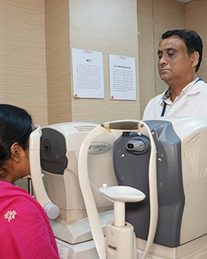
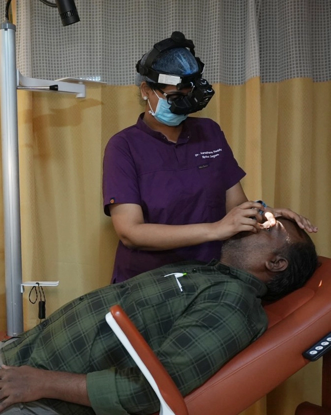
If you have diabetes, the most useful thing you can do for your eyes is dull: one dilated retina examination every year, whether or not your vision seems perfectly fine. Not a spectacle test. Not a pressure puff at an optical shop. A proper look at the back of both eyes, with drops, by someone who knows what early diabetic changes look like. Almost every case of severe diabetic vision loss we see could have been interrupted earlier, and the reason it was not is nearly always that the patient felt fine and did not come.
What high sugar does to the retina, and why you feel none of it
The retina is a thin sheet of nerve tissue lining the back of the eye, and it is fed by some of the smallest blood vessels in the body. Years of raised blood glucose damage the walls of those vessels. They begin to leak, and fluid and fat deposits collect in the retina. Some of them close off entirely, starving patches of tissue.
Starved retina then does something unhelpful. It sends out a chemical signal asking for new blood supply, and the eye obliges by growing fragile new vessels that were never meant to be there. These bleed into the cavity of the eye, and later contract and pull the retina off its base. That last stage, proliferative retinopathy with a tractional detachment, is the one that blinds people, and by the time it arrives the underlying process has been running quietly for years.
There is a second, more common route to lost vision: diabetic macular oedema, where fluid collects specifically at the macula, the small central patch you read with. This can happen at any stage, including quite early, and it is the reason a person with mild-looking retinopathy can still have genuinely blurred central vision.
The retina has no pain fibres. Vessels can leak, close and bleed without any sensation at all. Your central vision, the part you would notice, is a tiny fraction of the retinal surface, so a great deal can go wrong outside it while your reading stays sharp. And with two eyes, one covers for the other without your knowledge.
Blurred vision, floaters, or a sudden shower of black specks are real symptoms of diabetic eye disease, but they are late ones. Screening exists to find the disease in the years before those appear, when laser and injections work well and the retina is still healthy enough to save.
When to start, and how often
- Type 2 diabetes: get the first examination at the time of diagnosis. Type 2 is usually present for years before it is found, and a meaningful number of people already have retinopathy on the day they are told they are diabetic.
- Type 1 diabetes: start about five years after diagnosis, then annually.
- Children with diabetes: from around age ten or from puberty onwards, whichever comes first.
- Pregnancy: pregnancy can accelerate retinopathy sharply. If you are diabetic and planning a pregnancy, get examined before conceiving, then in each trimester. This does not apply to gestational diabetes that begins during the pregnancy itself.
- If changes are already present: every three to six months, as advised. Do not stretch these.
One more, and it catches people out: if your sugars have been very high for a long time and are then brought down rapidly, retinopathy can worsen temporarily before it settles. That is not a reason to control your diabetes any less tightly. It is a reason to have your eyes examined while you are doing it.
What the examination involves
Expect to be at the hospital for around two hours, most of it waiting for the drops to work. Someone should drive you home, or take an auto, because dilation leaves you glare-sensitive and unable to focus up close for four to six hours. Bring sunglasses.
The examination itself covers vision, eye pressure, the lens, and then a dilated view of the entire retina. Where needed we add an OCT scan, which measures retinal thickness at the macula in microns and picks up fluid long before it is visible to the eye, and ultra-widefield imaging on the Optos, which photographs a very large area of retina in a single capture and is useful for tracking change year on year. If leakage needs mapping precisely, an angiogram shows exactly which vessels are involved.
You should leave knowing which of five grades you fall into, from no retinopathy to proliferative disease, and when to return. If you are told only that it looks fine, ask what the grade was and whether the macula was scanned.
What treatment can and cannot do
Treatment is good at preserving vision and much less good at restoring it. That asymmetry is the whole argument for coming early.
- Laser photocoagulation treats the starved peripheral retina so it stops demanding new vessels. It works well and it is a trade: you sacrifice some peripheral and night vision to protect the centre.
- Anti-VEGF injections into the eye reduce leakage and macular swelling, and are the mainstay for macular oedema. They are given under anaesthetic drops and take a couple of minutes. The catch is that they are not a single event. A course commonly means several injections over the first year and continued review after. Cost varies a lot by which drug is used, from a few thousand rupees per injection for the least expensive option to several times that for the newer molecules, and your retina specialist should discuss that openly with you before starting rather than after.
- Vitrectomy surgery is for eyes that have bled heavily or where the retina is being pulled off. It is effective, and the vision it gives back depends on how much healthy retina is left to work with.
Two related points. Diabetics develop cataract earlier than others, and cataract surgery in a diabetic eye needs the retina assessed and stabilised first, which is a further reason to be under review. You can read what that involves on our cataract page. Diabetes also raises the risk of glaucoma, so pressure and optic disc are checked at the same visit.
The part no injection replaces
Nothing done inside the eye works as well as what is done outside it. Steady blood sugar over years, blood pressure kept controlled, cholesterol managed, and no smoking, together do more to protect your retina than any treatment we can offer once damage has started. Patients with well-controlled diabetes for twenty years frequently have retinas that look untouched. That is not luck.
If it has been more than a year since your last dilated examination, book one. Retina services run at Masab Tank, KPHB, Warangal, Nalgonda and Badvel, and there is more detail on our retina page. Bring your recent HbA1c report with you if you have one.